
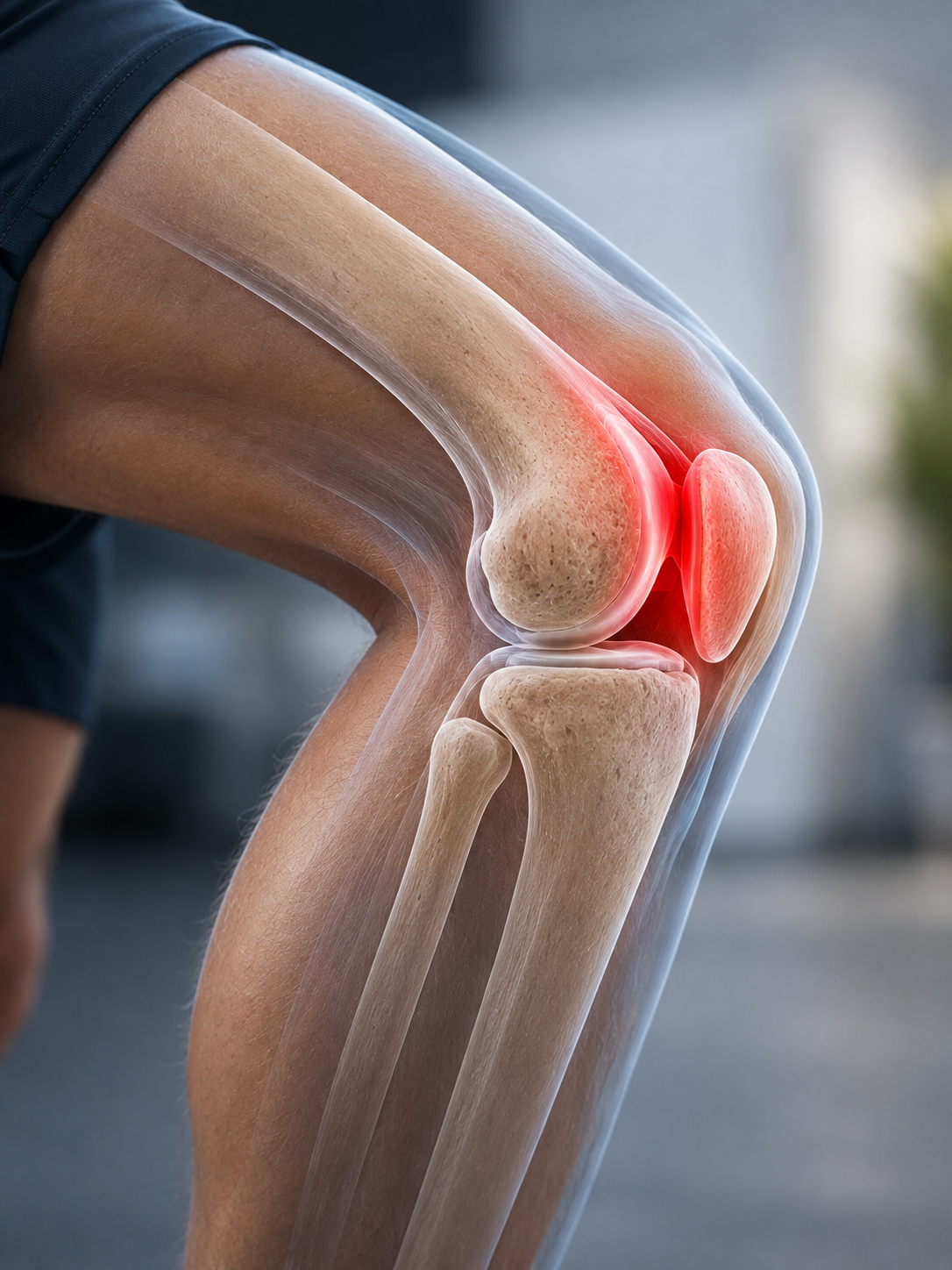
Patellofemoral (Kneecap) Problems: Pain Syndrome and Patellar Instability Explained
Pain around the front of the knee is one of the most common reasons people visit a physiotherapist, sports doctor or orthopaedic surgeon. Two conditions are frequently mentioned: patellofemoral pain syndrome (PFPS) and patellar instability. Although they both involve the kneecap (patella), they are very different problems and often require different treatments.
Patellofemoral pain syndrome usually causes pain around or behind the kneecap without the kneecap actually coming out of place. It is often related to overuse, training errors or changes in the way forces pass through the front of the knee. Patellar instability, on the other hand, occurs when the kneecap partially slips out of its groove (subluxation) or completely dislocates, usually after a twisting injury or because of underlying anatomical factors.
Understanding the difference is important because treatment depends on making the correct diagnosis. Many people improve with targeted physiotherapy and rehabilitation, while others — particularly those with recurrent instability — may benefit from surgery to restore stability.
This guide explains how the patellofemoral joint works, why these conditions develop, how they differ and the treatments that are most likely to help.
Key points
- Patellofemoral pain syndrome and patellar instability are different conditions, although both affect the kneecap.
- Patellofemoral pain syndrome usually causes pain around the front of the knee without the kneecap dislocating.
- Patellar instability occurs when the kneecap repeatedly slips or dislocates from its normal position.
- Most people with patellofemoral pain improve with progressive strengthening and physiotherapy.
- Surgery is rarely needed for patellofemoral pain syndrome but may be appropriate for recurrent patellar instability or after certain first-time dislocations with associated injuries.
Understanding the patellofemoral joint
To understand these conditions, it helps to know how the kneecap normally works.
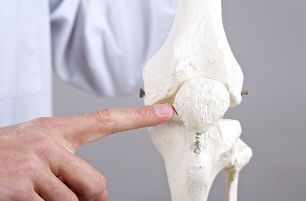
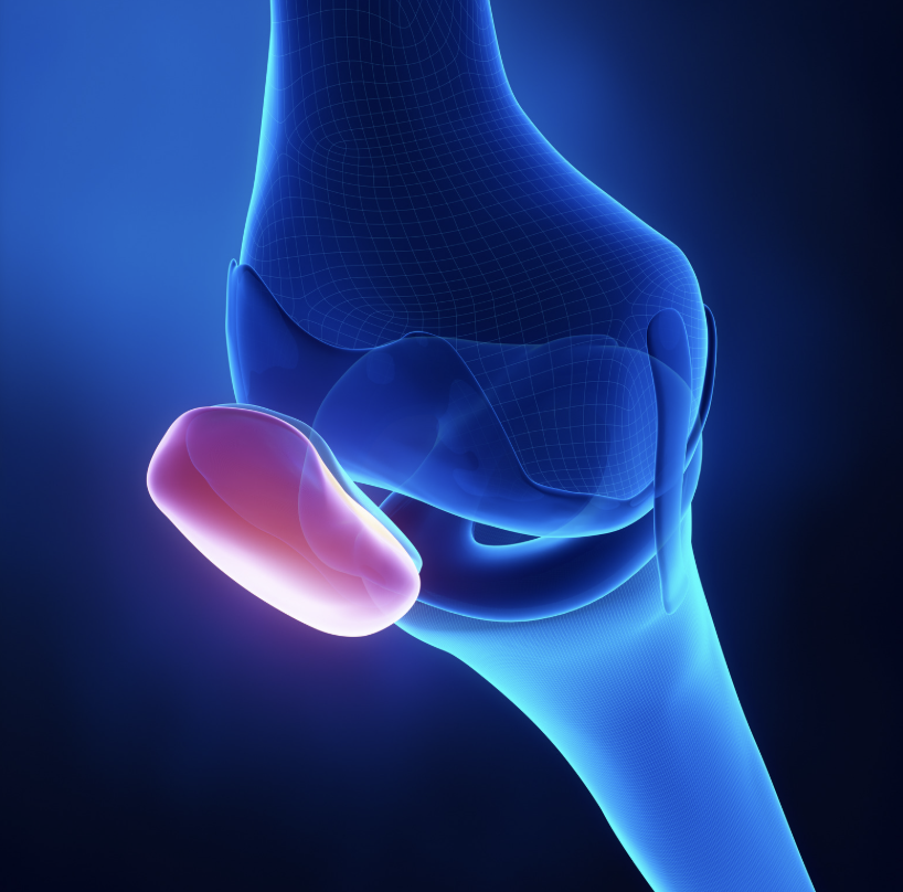
The patella is the small bone at the front of the knee. Rather than simply covering the joint, it acts like a pulley within the quadriceps tendon, increasing the efficiency of the thigh muscles when straightening the knee.
As the knee bends and straightens, the kneecap glides within a groove at the end of the thigh bone called the trochlear groove.
Several structures work together to keep the kneecap centred within this groove: the shape and depth of the trochlear groove, the quadriceps muscles, the patellar tendon, the medial patellofemoral ligament (MPFL), the surrounding soft tissues, and the overall alignment of the leg.
When these structures work together, the kneecap moves smoothly throughout knee movement. When they do not, problems can develop.
What does the patella actually do?
Many people assume the kneecap simply protects the front of the knee. In reality, it performs several important functions. It increases the power of the quadriceps muscles, improves the efficiency of knee extension, reduces stress on the quadriceps tendon, helps distribute forces across the front of the knee, and protects the knee joint during kneeling and direct impact.
During activities such as climbing stairs, squatting or getting up from a chair, the forces passing through the patellofemoral joint can reach several times body weight. This explains why problems affecting the kneecap often become painful during these activities.
Patellofemoral pain syndrome vs patellar instability
Although these conditions are often confused, they are fundamentally different.
| Patellofemoral pain syndrome (PFPS) | Patellar instability |
|---|---|
| Pain around or behind the kneecap | Kneecap partially or completely slips out of place |
| Usually develops gradually | Often follows a twisting injury or previous dislocation |
| Kneecap remains within the groove | Kneecap leaves the groove (temporarily or completely) |
| Pain is the main symptom | Instability is the main symptom |
| Usually no major swelling | Significant swelling is common after a dislocation |
| MRI often normal or shows mild cartilage changes | MRI may show MPFL injury, bone bruising or cartilage damage |
| Usually treated with physiotherapy | Treatment depends on the severity and recurrence of instability |
| Surgery is uncommon | Surgery may be recommended in selected patients |
The important point is that pain does not necessarily mean instability, and instability does not always cause ongoing pain between episodes. Correct diagnosis is therefore essential before deciding on treatment.
What is patellofemoral pain syndrome?
Patellofemoral pain syndrome (PFPS) describes pain arising from the front of the knee, usually around or behind the kneecap, without another major structural injury explaining the symptoms. It is one of the most common causes of knee pain in runners, cyclists, footballers, young adults, teenagers, people increasing their exercise levels and individuals returning to sport after injury.
Despite being common, PFPS is often misunderstood. For many years it was thought to occur because the kneecap was “tracking incorrectly”. We now know that the picture is more complex.
Rather than being caused by one single abnormality, patellofemoral pain usually develops because the forces passing through the front of the knee become greater than the tissues can comfortably tolerate. This is sometimes described as a load versus capacity problem.
If the demands placed on the knee suddenly increase — or the muscles supporting the knee become weaker — the tissues around the kneecap may become painful even though no major injury has occurred. This also explains why strengthening exercises are often so effective.
Why does patellofemoral pain develop?
There is rarely one single cause. Instead, several factors often combine together. These may include sudden increases in running or sporting activity, weak quadriceps muscles, weakness of the hip muscles, reduced control of leg movement, altered movement patterns, prolonged sitting with bent knees, previous injury, reduced flexibility, muscle imbalance and occasionally foot biomechanics.
Importantly, having one of these factors does not automatically mean you will develop pain. Most people develop symptoms because several small factors occur together.
Is my kneecap actually damaged?
Usually not. Many people worry that pain around the kneecap means the cartilage is wearing away. Fortunately, this is rarely the case. In many people with patellofemoral pain, X-rays are normal, MRI scans are normal, there is no major cartilage injury, the ligaments are intact and the kneecap remains stable.
Instead, the pain is thought to arise from sensitive tissues around the patellofemoral joint responding to increased loading. This is reassuring because it means many people recover without surgery.
Why does it hurt going down stairs?
One of the classic symptoms of patellofemoral pain is discomfort when walking downstairs, squatting, kneeling or getting out of a low chair.
This happens because these activities place particularly high loads across the patellofemoral joint. As the knee bends, the contact force between the kneecap and the femur increases. If the surrounding tissues have become sensitive, these activities often reproduce symptoms. The same principle explains why prolonged sitting with bent knees — sometimes called “movie-goer's knee” — can become uncomfortable.
Patellofemoral pain does not mean you should stop exercising
Many people understandably avoid activity once pain begins. Unfortunately, complete rest is rarely the answer. Avoiding all activity can lead to weaker quadriceps muscles, weaker hip muscles, reduced fitness and poorer movement control. Over time, this can actually make symptoms more persistent.
Instead, treatment usually focuses on modifying activity rather than stopping it completely. The aim is to reduce pain while gradually rebuilding the strength and capacity of the knee so it can tolerate higher levels of activity again.
Symptoms of patellofemoral pain syndrome
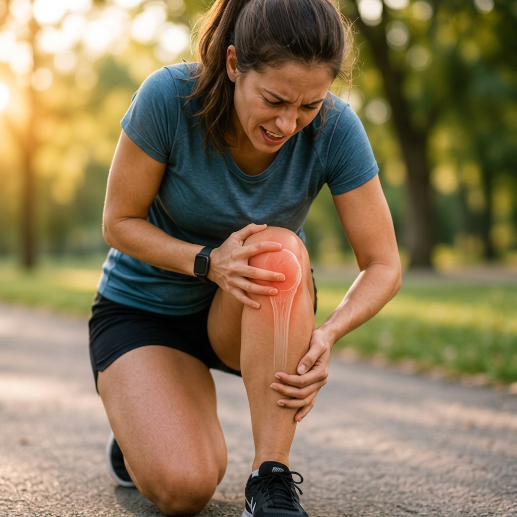
Patellofemoral pain syndrome usually develops gradually rather than after one specific injury. People often describe an aching pain around or behind the kneecap that becomes worse during activities that increase pressure across the front of the knee.
Typical symptoms include pain around or behind the kneecap; pain when walking downstairs; pain during squatting or lunging; discomfort when running, particularly downhill; pain after prolonged sitting (“movie-goer's knee”); pain when kneeling; stiffness after sitting for long periods; occasional clicking or grinding sensations; and difficulty returning to sport.
Unlike ligament injuries, the knee usually does not feel unstable, and the kneecap does not actually come out of joint.
Many patients notice that symptoms fluctuate. They may improve for several weeks before returning after increasing activity, changing training intensity or spending more time on their feet.
What is patellar instability?
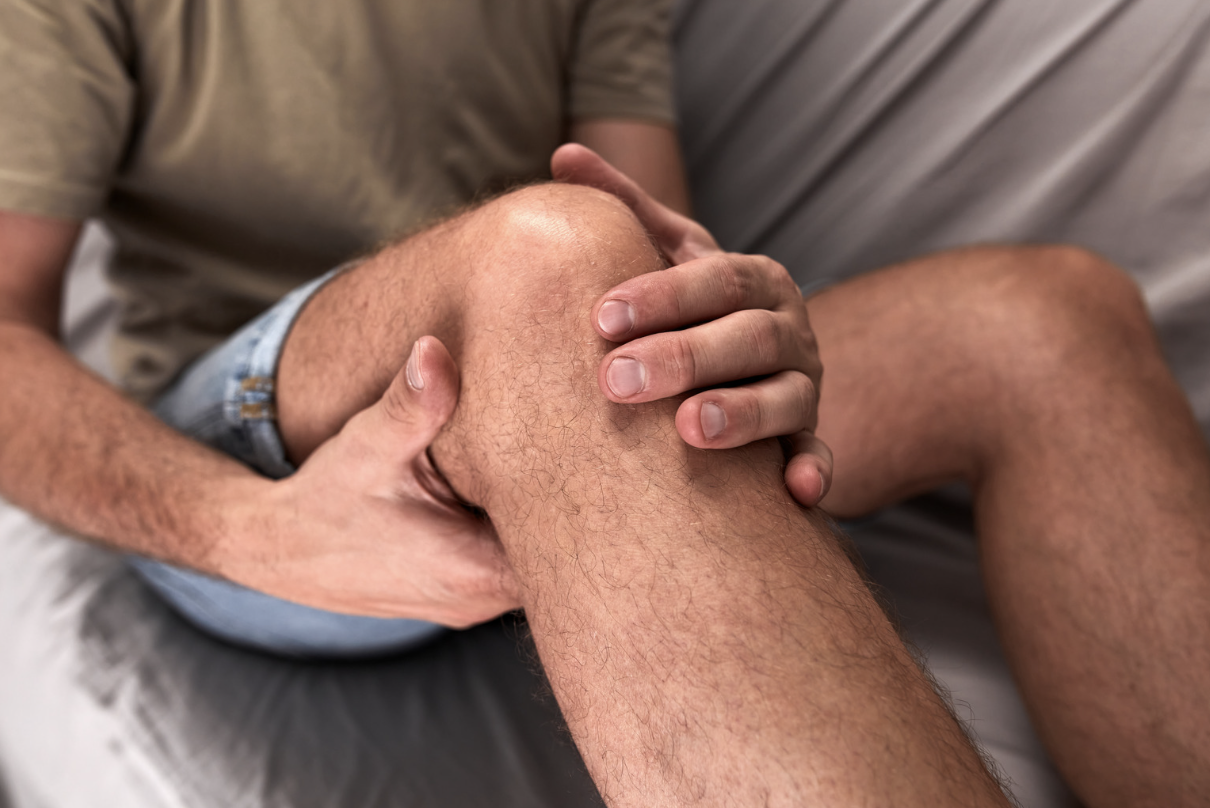
Patellar instability is a completely different condition. Instead of simply becoming painful, the kneecap actually moves too far out of its normal groove.
This may happen once after an injury, occasionally during sport, or repeatedly over many years.
Some people describe the kneecap as “slipping”. Others say “it popped out”, or “my knee gave way”. These descriptions are often clues that instability, rather than simple patellofemoral pain, is present.
Subluxation vs dislocation
Patellar subluxation
A subluxation occurs when the kneecap partially slips out of its groove before moving back into place by itself. The person may experience sudden pain, a feeling that the kneecap shifted, temporary loss of confidence and swelling afterwards.
Some people only experience this once. Others experience repeated subluxations over many years.
Patellar dislocation
A dislocation occurs when the kneecap completely leaves its normal groove. Most dislocations occur towards the outside (lateral side) of the knee.
The knee usually becomes immediately painful, swollen, difficult to bend, difficult to straighten and difficult to walk on. Sometimes the kneecap slides back into place on its own. Sometimes it remains dislocated until it is relocated in the Emergency Department.
What happens during a patellar dislocation?
When the kneecap dislocates, several structures can be injured at the same time. These may include the medial patellofemoral ligament (MPFL), joint cartilage, underlying bone and surrounding soft tissues.
The MPFL is the main ligament preventing the kneecap from moving too far towards the outside of the knee during the first 20–30 degrees of bending. It acts rather like a seatbelt. When the kneecap dislocates, this ligament is commonly torn.
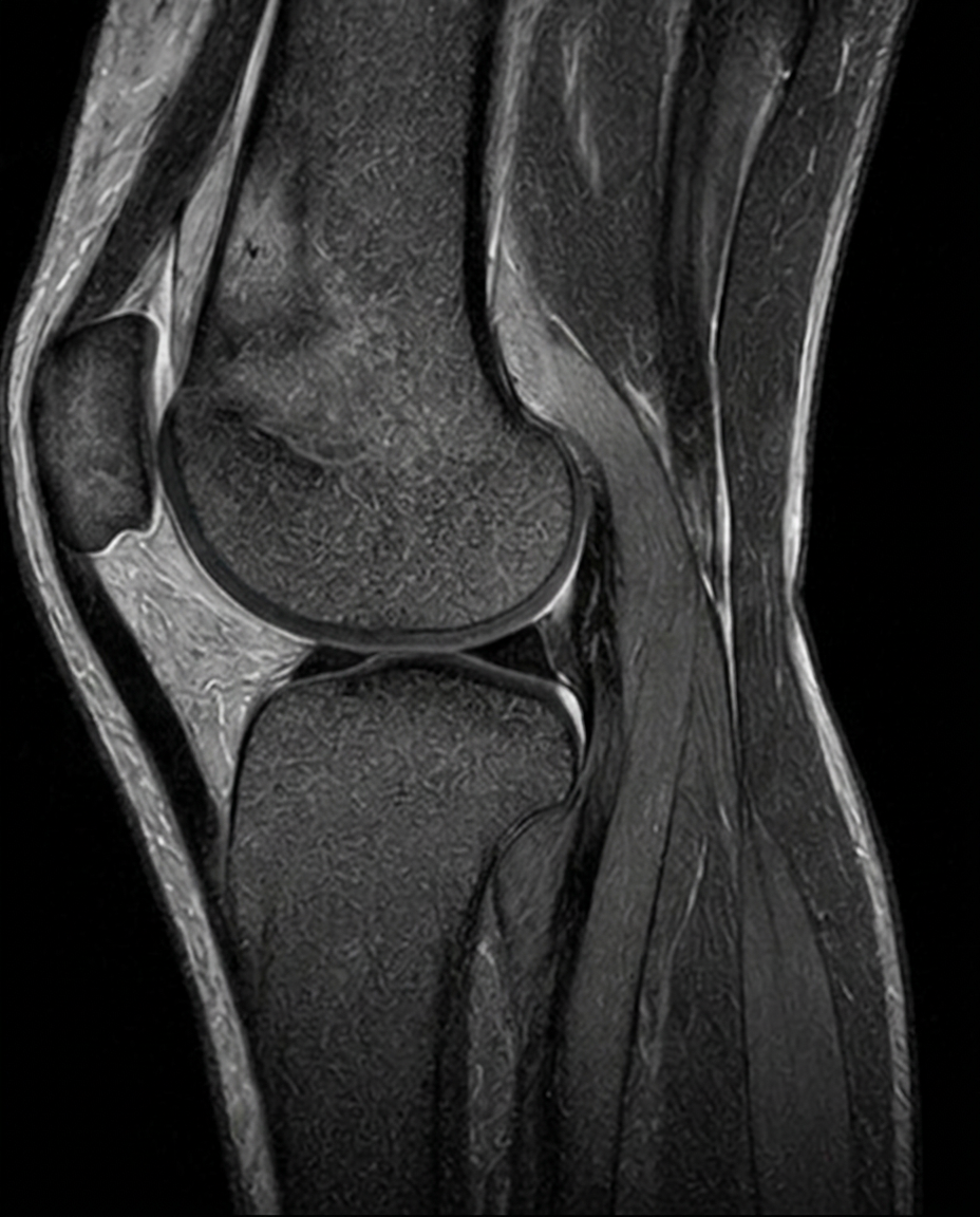
In addition to ligament injury, the kneecap and the outer part of the femur may strike each other with considerable force. This can cause bone bruising, cartilage injury and small fractures involving cartilage and bone (osteochondral fractures).
This is one reason why MRI is often recommended after a first-time dislocation, particularly in young patients, as it helps identify injuries that may influence treatment. The BOAST guidance recommends MRI following first-time dislocation when an osteochondral injury is suspected or when further assessment is needed.
Why do some people keep dislocating their kneecap?
Many people experience one dislocation and never have another. Others develop recurrent instability. Why? Usually because they have one or more anatomical factors that make the kneecap less stable.
Trochlear dysplasia
Normally, the kneecap sits within a well-defined groove. In some people this groove is naturally shallow. This is called trochlear dysplasia. Imagine balancing a golf ball in a deep egg cup, versus balancing it on a flat saucer. The ball is far more likely to roll off the saucer. The same principle applies to the kneecap.
Trochlear dysplasia is one of the strongest risk factors for recurrent instability.
Patella alta
Some people have a kneecap that naturally sits higher than average. This is known as patella alta. Because the kneecap starts higher, it engages the groove later as the knee bends. During this early part of movement there is less bony stability, making dislocation more likely.
Increased TT–TG distance
The tibial tubercle is the point where the patellar tendon attaches to the shin bone. If this attachment lies further towards the outside than normal, the pull of the quadriceps tends to draw the kneecap laterally. This is measured on CT or MRI as the tibial tubercle–trochlear groove (TT–TG) distance. A markedly increased TT–TG distance increases the risk of recurrent instability and may influence surgical planning in selected patients.
Generalised joint hypermobility
Some people naturally have looser ligaments throughout their body. They may also notice very flexible fingers, elbows that straighten beyond normal, hypermobile shoulders and ankles that repeatedly sprain.
Looser soft tissues provide less restraint to the kneecap, increasing the chance of recurrent instability.
Previous dislocation
The strongest predictor of another dislocation is having already experienced one. This is partly because the MPFL often heals in a lengthened position after injury. Without its normal restraint, the kneecap may slip more easily during twisting movements.
Muscle weakness
Although muscle weakness alone rarely causes dislocation, weak quadriceps and hip muscles reduce dynamic control of the knee. Strengthening these muscles forms an important part of rehabilitation after both first-time and recurrent instability.
Risk factors for patellofemoral pain syndrome
Unlike instability, patellofemoral pain is usually not caused by one single anatomical abnormality. Instead, several factors combine together. These include sudden increases in training, poor rehabilitation after injury, weak quadriceps, weak hip muscles, reduced control of the pelvis during running, prolonged sitting, repetitive stair climbing, running downhill, reduced flexibility and previous episodes of knee pain.
The important point is that having one of these factors does not guarantee you will develop pain. Rather, symptoms occur when the load placed upon the knee exceeds what the tissues can comfortably tolerate.
How are these conditions diagnosed?
Diagnosis begins with listening carefully to your history. Often, the story itself provides the biggest clue.
Patellofemoral pain syndrome
Typically develops gradually. Pain is the main symptom. Activities such as stairs, squatting, kneeling and prolonged sitting usually reproduce symptoms. The kneecap has never actually come out of place.
Patellar instability
Often follows a twisting injury, sporting injury or awkward landing. The patient frequently remembers “my kneecap came out” or “it popped sideways”. There is often immediate swelling.
Many patients become anxious about repeating twisting movements because they fear another dislocation.
Physical examination
Your clinician will assess your walking pattern, knee alignment, swelling, tenderness, range of movement, quadriceps strength, hip strength, flexibility and ligament stability.
Patellar apprehension test
The examiner gently moves the kneecap towards the outside of the knee. Patients with instability often become anxious or instinctively tighten their muscles because they feel the kneecap may dislocate again. This differs from simple patellofemoral pain, where discomfort may be present but the sensation of impending dislocation is absent.
Single-leg squat
Watching someone squat on one leg provides valuable information about hip control, knee alignment, balance and movement patterns. Poor movement control often contributes to patellofemoral pain and helps guide physiotherapy.
Do you need an X-ray?
After a first-time dislocation, X-rays are usually recommended. They help identify fractures, osteochondral fragments, alignment and obvious anatomical abnormalities.
For patellofemoral pain syndrome, X-rays are often normal and are not always necessary, particularly in younger patients with a typical history and examination.
When is an MRI needed?
MRI provides much more information about the soft tissues than an X-ray. It can identify MPFL tears, cartilage injuries, bone bruising, loose bodies, osteochondral fractures and other ligament injuries.
Following a first-time dislocation, MRI is commonly performed because identifying a significant cartilage injury or loose fragment may change treatment.
For uncomplicated patellofemoral pain syndrome, MRI is often not required initially. Many people improve with physiotherapy without ever needing advanced imaging.
Is a CT scan ever needed?
Sometimes. CT scans are particularly useful when detailed assessment of bone anatomy is required. They may be used to assess trochlear dysplasia, TT–TG distance, rotational alignment and surgical planning.
CT is therefore more commonly used when recurrent instability is being investigated rather than routine patellofemoral pain.
When should you seek urgent medical advice?
You should seek prompt assessment if your kneecap has dislocated for the first time; the knee remains obviously out of joint; you cannot straighten the knee; there is a large swelling immediately after injury; you cannot bear weight; the knee repeatedly gives way; you develop numbness in the foot; or you suspect a fracture.
A first-time patellar dislocation should not simply be dismissed as “a sprain”. Early assessment helps identify associated injuries and determine whether further imaging or specialist referral is needed.
Treatment of patellofemoral pain syndrome
One of the most reassuring things about patellofemoral pain syndrome is that the vast majority of people recover without surgery. Although symptoms can sometimes be frustrating and slow to settle, most people improve significantly with the right rehabilitation programme.
The aim of treatment is not simply to reduce pain today. It is to improve the knee's ability to cope with everyday life, exercise and sport over the long term. Rather than trying to “fix” one specific structure, treatment focuses on gradually increasing the capacity of the entire lower limb.
For most patients, this means combining education, activity modification, progressive strengthening, movement retraining and a gradual return to sport.
Why exercise is the most important treatment
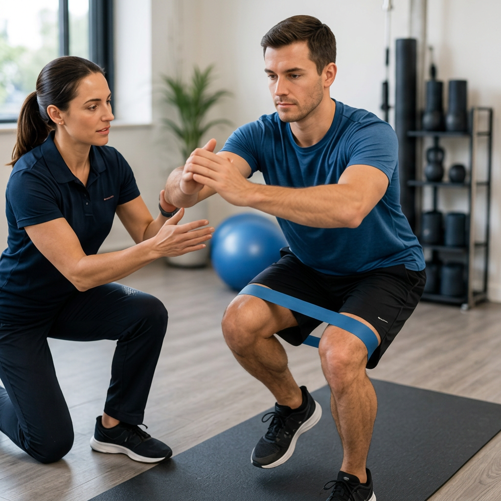
Many people hope there is a quick solution — a brace, injection or operation. Unfortunately, there is no treatment that works as well as a properly designed rehabilitation programme. Research consistently shows that exercise therapy is the cornerstone of treatment for patellofemoral pain syndrome.
Exercise works because it improves muscle strength, improves movement control, reduces stress on the patellofemoral joint, increases tissue tolerance, restores confidence and allows a gradual return to activity.
Importantly, exercise is not simply about strengthening the quadriceps. Modern rehabilitation looks at the whole lower limb.
Understanding “load versus capacity”
One of the easiest ways to understand patellofemoral pain is to imagine a bridge. Every bridge has a maximum weight it can safely support. If traffic gradually increases beyond that capacity, damage begins to occur.
Your knee works in much the same way. Every day the patellofemoral joint experiences load from walking, climbing stairs, running, jumping, squatting and kneeling.
Normally the muscles and surrounding tissues cope easily. However, if training suddenly increases, muscles become weaker, activity changes dramatically or recovery is inadequate, the load placed on the knee may exceed what the tissues can comfortably tolerate. The result is pain.
The solution is therefore not complete rest. It is gradually increasing the knee's capacity so it can once again tolerate these activities.
Should you stop exercising?
Usually not. Many people stop all exercise as soon as knee pain develops. Unfortunately this often makes recovery slower. Complete rest leads to weaker muscles, reduced fitness, poorer movement control, greater stiffness and lower confidence.
Instead, the goal is relative rest. This means reducing activities that cause significant pain while continuing to stay active. For example, instead of running 10 km every day, you may temporarily reduce to shorter runs, cycling, swimming, walking or cross-training. As symptoms improve, running can gradually be reintroduced.
Strengthening the quadriceps
The quadriceps are the largest muscles at the front of the thigh. They play a major role in controlling the kneecap during movement. Weakness or poor activation may increase the stress placed on the patellofemoral joint.
Early exercises often include quadriceps contractions, straight-leg raises, mini squats, wall sits and sit-to-stands.
As strength improves, exercises become progressively harder. Later stages may include split squats, step-ups, Bulgarian squats, leg press and single-leg squats.
The aim is not simply to build muscle. It is to improve how the knee functions during everyday movement.
Hip strength is just as important
One of the biggest changes in rehabilitation over the past decade has been recognising the importance of the hip muscles. The gluteal muscles help control pelvic position, thigh rotation and knee alignment.
When these muscles are weak, the knee may move inwards during activities such as running, landing, stair climbing and squatting. This can increase stress across the patellofemoral joint.
Hip exercises commonly include bridges, clamshells, side leg raises, monster walks, resisted side stepping and single-leg balance exercises. Many patients notice significant improvement once both the hip and quadriceps are strengthened together.
Improving movement patterns
Sometimes strength is not the only issue. The way someone moves may also contribute to symptoms. A physiotherapist may assess running technique, jumping mechanics, landing control, single-leg squat, stair climbing and balance.
Small changes in movement can sometimes reduce the load passing through the front of the knee. Examples include improving cadence while running, avoiding excessive inward knee movement, improving trunk control and strengthening the core. These changes should always be individualised rather than applied to everyone.
Returning to running
One of the most common questions is: “When can I start running again?” The answer depends on symptoms rather than the calendar.
Before returning to running you should usually have minimal pain during walking, full knee movement, good quadriceps strength, good hip strength and excellent single-leg control.
Running should then increase gradually. A typical progression is: walking → walk-run intervals → continuous running → longer distances → hill running → speed work → sport-specific drills.
Increasing too quickly is one of the commonest reasons symptoms return.
Is pain during exercise acceptable?
Many patients worry that any pain means they are causing damage. Fortunately this is usually not true. Some discomfort during rehabilitation is common.
A useful guide is: mild discomfort during exercise is often acceptable; symptoms should settle reasonably quickly afterwards; and pain should not continue increasing day after day.
If pain becomes progressively worse or lasts for several days after exercise, the programme may simply need adjusting. This does not usually mean you have injured the knee again.
Does taping help?
Patellar taping is sometimes used during rehabilitation. It may reduce pain, improve confidence and allow exercises to be performed more comfortably.
However, taping is not a cure. Its main purpose is to make rehabilitation easier while strength improves. Many physiotherapists use taping only during the early stages of treatment.
What about knee braces?
Many knee braces are advertised for patellofemoral pain. Some people feel they provide support, confidence and temporary symptom relief.
However, braces do not strengthen muscles or correct the underlying problem. For this reason they should generally be viewed as an addition to rehabilitation rather than a replacement for exercise.
Can foot orthotics help?
Some people with excessive foot pronation (rolling inwards) may benefit from foot orthoses. These aim to improve lower-limb alignment during walking and running.
However, they do not help everyone, they are not routinely required, and they work best when combined with strengthening exercises. They should be prescribed based on individual assessment rather than bought routinely.
Pain relief
Medication can help reduce symptoms during rehabilitation. However, painkillers do not treat the underlying cause. Simple options include paracetamol, topical anti-inflammatory gels and oral anti-inflammatory medication when appropriate.
Medication should be viewed as a way of allowing rehabilitation to continue rather than as the primary treatment.
Do injections work?
Patients often ask whether an injection could solve the problem. Unlike arthritis, there is very little evidence supporting injections for typical patellofemoral pain syndrome. Steroid injections are therefore not routinely recommended for uncomplicated PFPS.
Occasionally injections may be considered if another condition exists alongside patellofemoral pain, such as inflammation around a tendon or other pathology, but they are not part of standard treatment for isolated PFPS.
Is surgery ever needed for patellofemoral pain?
The reassuring answer is: very rarely. Patellofemoral pain syndrome is usually not a surgical disease. Operations aimed purely at treating anterior knee pain have generally produced inconsistent results.
If someone has normal stability, no major cartilage injury and no significant anatomical abnormality, then surgery is unlikely to provide better results than well-designed rehabilitation. This is why surgeons usually recommend a comprehensive physiotherapy programme before considering any operation.
How long does recovery take?
Recovery varies considerably. Some people improve within a few weeks. Others require several months.
| Time | Expected progress |
|---|---|
| 2–6 weeks | Pain begins to settle, confidence improves |
| 6–12 weeks | Strength increases, daily activities become easier |
| 3–6 months | Return to running and sport for many patients |
| 6–12 months | Continued improvements in strength, endurance and performance |
The important point is that improvement is rarely completely linear. Most people experience occasional flare-ups along the way. These do not necessarily mean rehabilitation has failed.
What happens if you ignore it?
Continuing to push through significant pain without modifying activity may lead to persistent symptoms, reduced sporting performance, muscle weakness, frustration and prolonged recovery.
Fortunately, patellofemoral pain syndrome does not usually cause rapid joint damage, but chronic pain can become more difficult to treat if it is ignored for long periods. Early assessment and a structured rehabilitation programme generally provide the best chance of recovery.
Treatment of patellar instability
Unlike patellofemoral pain syndrome, patellar instability is a condition where the kneecap actually slips out of its normal groove. The treatment pathway is therefore very different.
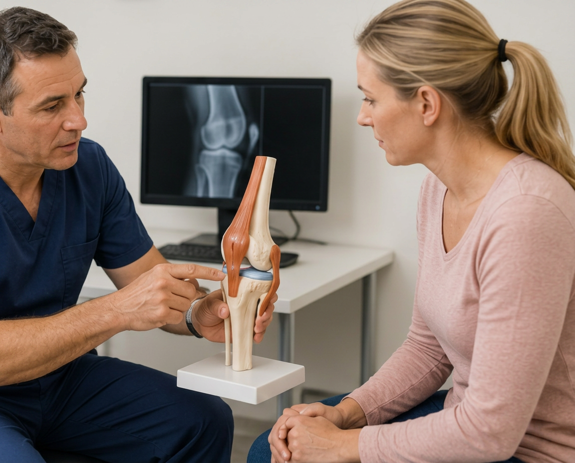
The first question your orthopaedic surgeon will ask is not “have you dislocated your kneecap?” Instead, it is “why did it dislocate?” Understanding why the kneecap became unstable is the key to choosing the correct treatment.
Some people suffer a single traumatic dislocation and never experience another episode. Others have underlying anatomical factors that make repeated dislocations much more likely.
The aim of treatment is not simply to prevent another dislocation — it is also to protect the cartilage of the patellofemoral joint and reduce the risk of developing arthritis later in life.
Treatment after a first-time patellar dislocation
A first-time dislocation is frightening. The knee often swells rapidly, walking becomes difficult and many people worry they have torn a major ligament such as the ACL.
Fortunately, most first-time patellar dislocations do not require surgery. Current UK guidance recommends that most patients are managed initially with non-operative treatment, while ensuring that important associated injuries — particularly osteochondral fractures — are not missed.
Initial treatment usually includes ice, compression, elevation, appropriate pain relief, a supportive brace if required, early restoration of movement, physiotherapy and MRI assessment when indicated.
The older practice of placing the knee in a rigid plaster cast for several weeks has largely been abandoned because prolonged immobilisation delays recovery and leads to muscle wasting and stiffness.
Why is an MRI important?
Following a first dislocation, an MRI scan is often one of the most useful investigations. It helps identify MPFL tears, cartilage injuries, bone bruising, loose fragments of cartilage or bone, osteochondral fractures and associated ligament injuries.
This information is important because a loose fragment inside the knee may require early surgery, whereas an isolated MPFL injury without major cartilage damage can often be treated non-operatively.
Physiotherapy after a dislocation
Once pain and swelling begin to settle, rehabilitation becomes the most important part of recovery. The goals are to reduce swelling, regain full movement, restore quadriceps strength, improve hip strength, improve balance, rebuild confidence and gradually return to sport.
Early exercises may include quadriceps activation, straight-leg raises, heel slides, gentle knee bending and calf pumps.
As recovery progresses, rehabilitation becomes more demanding. Later exercises include squats, lunges, step-ups, single-leg balance, hopping drills, agility exercises and sport-specific training.
The programme should be individualised and progressed gradually rather than following a fixed timetable.
Will my kneecap dislocate again?
This is the question almost everyone asks. Unfortunately, there is no simple yes or no answer. Some people never experience another episode. Others continue to have recurrent instability.
The risk of recurrence depends on several factors, including age, sex, trochlear dysplasia, patella alta, generalised hypermobility, increased TT–TG distance, family history and previous dislocations.
Younger patients, particularly teenagers and those with significant anatomical abnormalities, have the highest risk of recurrent instability. Published series report recurrence rates after a first-time dislocation ranging from around 20% to as high as 80% in high-risk groups.
When is surgery recommended?
Most patients do not require surgery after a first dislocation. However, surgery becomes more likely if there is a large osteochondral fracture, there is a loose fragment within the knee, the kneecap continues to dislocate, recurrent instability affects daily life or sport, or significant anatomical abnormalities increase the risk of recurrence.
Current British guidance generally supports reserving stabilisation surgery for recurrent instability rather than routine surgery after every first dislocation. The decision is always individual and based on both the patient's symptoms and the underlying anatomy.
MPFL reconstruction
The medial patellofemoral ligament (MPFL) is the main soft-tissue restraint preventing the kneecap from moving too far towards the outside of the knee during the early stages of knee bending. Because this ligament is commonly torn during a dislocation, reconstruction is one of the most frequently performed operations for recurrent instability.
During surgery, a tendon graft is used to create a new MPFL, the graft is attached to the patella and femur, and the new ligament restores stability while still allowing normal movement.
The aim is not to make the kneecap rigid. Instead, it restores the normal restraint that was lost during injury. MPFL reconstruction produces excellent results in carefully selected patients but is not appropriate for everyone.
Tibial tubercle osteotomy
Some patients have an abnormal alignment of the patellar tendon. Rather than simply reconstructing the ligament, the underlying pull of the tendon also needs correcting.
This operation involves moving the tibial tubercle — the bony attachment of the patellar tendon — to improve the alignment of the kneecap. Depending on the individual's anatomy, this can reduce the sideways pull on the kneecap, improve tracking, lower a high-riding patella (patella alta) and reduce the risk of further dislocation.
Because this is a bone operation, it is generally reserved for patients with significant anatomical abnormalities and is not usually performed in children with open growth plates.
Trochleoplasty
In a small number of patients, the trochlear groove itself is extremely shallow. In these cases, simply reconstructing the MPFL may not provide sufficient stability. A trochleoplasty reshapes the groove so that the kneecap sits more securely. This is a specialised procedure performed in selected patients after careful assessment and is usually reserved for severe trochlear dysplasia.
Can more than one procedure be needed?
Yes. Patellar instability is rarely caused by a single abnormality. For example, a patient may have trochlear dysplasia, patella alta, a torn MPFL and an increased TT–TG distance. Treating only one of these problems may leave the kneecap unstable. This is why careful pre-operative planning is so important.
Some patients benefit from MPFL reconstruction alone, MPFL reconstruction combined with tibial tubercle osteotomy, cartilage repair at the same operation, or other alignment procedures depending on the individual's anatomy.
Modern surgery aims to correct the factors responsible for instability rather than applying the same operation to everyone.
Why isolated lateral release is no longer recommended
Years ago, many patients underwent an operation called an isolated lateral release, in which tight tissues on the outside of the kneecap were divided.
We now know that this operation does not treat the underlying causes of patellar instability and can even make instability worse in some patients. For this reason, isolated lateral release has no role as a treatment for patellar instability.
Recovery after patellar stabilisation surgery
Recovery depends on the operation performed.
| Stage | What to expect |
|---|---|
| First 2 weeks | Swelling control, wound care, regaining knee extension, quadriceps activation, walking with crutches if required |
| 2–6 weeks | Improving knee bending, progressive strengthening, reducing walking aids, improving confidence |
| 6–12 weeks | Advanced strengthening, balance exercises, cycling, swimming, functional rehabilitation |
| 3–6 months | Jogging, hopping, agility drills, sport-specific exercises |
| 6–9 months | Return to pivoting sports once strength, balance and movement control are restored and return-to-sport testing is completed |
Long-term outlook
The outlook for most patients is very good. Many people return to football, rugby, netball, skiing, tennis and running.
The earlier instability is recognised and appropriately treated, the better the opportunity to protect the cartilage within the patellofemoral joint.
Repeated dislocations can damage the cartilage on both the kneecap and the femur, increasing the risk of developing patellofemoral arthritis later in life. Preventing recurrent instability is therefore about preserving the long-term health of the knee as well as restoring confidence and function.
Frequently asked questions
Will I always need surgery if my kneecap dislocates?
No. Most first-time dislocations are managed without surgery unless there is a significant cartilage injury, loose fragment or another indication for early operative treatment.
Can physiotherapy stop my kneecap dislocating?
Physiotherapy improves strength, movement control and confidence and is an essential part of treatment. However, it cannot change structural factors such as a shallow trochlear groove or patella alta. In some people, surgery is needed to address these anatomical problems.
Will I develop arthritis?
Not everyone does, but repeated instability episodes increase the risk because each dislocation can damage the joint cartilage.
Can I return to sport?
Most patients can return to sport after appropriate rehabilitation. The timing depends on restoring strength, stability, balance and confidence rather than simply reaching a certain number of weeks after injury or surgery.
The OrthoZone take-home message
Patellofemoral pain syndrome and patellar instability may both affect the kneecap, but they are very different conditions. Patellofemoral pain is usually managed successfully with education, activity modification and progressive strengthening, while patellar instability requires careful assessment to identify why the kneecap has become unstable. Most first-time dislocations can be treated without surgery, but recurrent instability, significant cartilage injury or certain anatomical abnormalities may require specialist surgical treatment. The key to achieving the best outcome is making the correct diagnosis and choosing treatment that is tailored to the individual rather than applying the same approach to every patient.
Top tips
- A first-time patellar dislocation does not automatically mean you need surgery. Most patients recover well with appropriate rehabilitation, but imaging is important to exclude associated cartilage injuries.
- Don't ignore repeated episodes of instability. Every dislocation has the potential to damage the cartilage lining the joint.
- Rehabilitation matters. Restoring quadriceps and hip strength, balance and movement control is essential whether or not surgery is required.
- Surgery should be tailored to the individual. The best operation depends on why the kneecap is unstable, not simply on the fact that it has dislocated.
- The goal is long-term knee health. Successful treatment aims to restore stability, protect the cartilage and help you return confidently to work, sport and everyday life.
Related articles
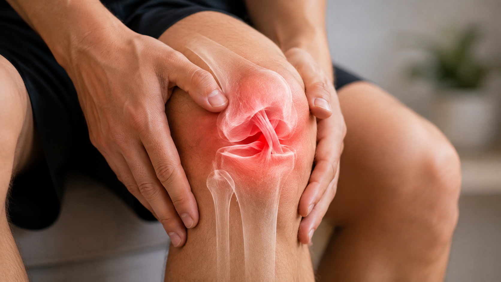
ACL Injury: Symptoms, Treatment, Surgery and Recovery
An evidence-based guide to ACL injuries: symptoms, diagnosis, rehabilitation, when surgery is needed, graft choices and return to sport.
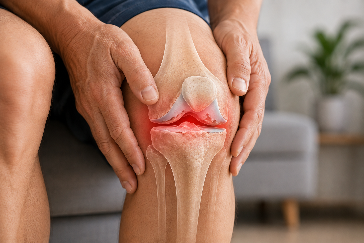
Knee Arthritis (Osteoarthritis): Symptoms, Causes, Diagnosis and Treatment
An evidence-based guide to knee osteoarthritis: symptoms, causes, diagnosis, exercise, weight management, injections and when knee replacement is recommended.
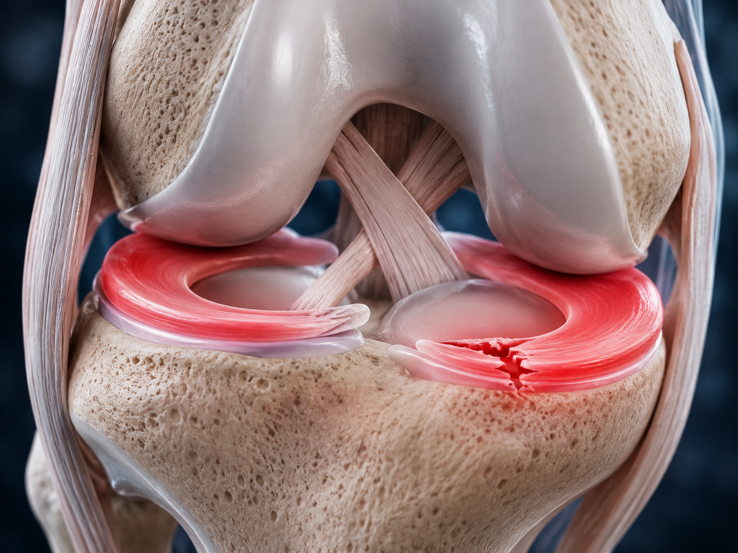
Meniscus Tears: Symptoms, Treatment, Surgery and Recovery
An evidence-based guide to meniscus tears: symptoms, diagnosis, physiotherapy, meniscal repair, keyhole surgery and recovery.