
Shoulder Dislocation and Instability: Treatment, Surgery and Recovery
A shoulder dislocation happens when the ball at the top of the upper arm comes completely out of its socket. It is usually a painful and frightening injury, but the immediate treatment — putting the shoulder back into place — is only the first part of the story.
The next question is whether the shoulder will remain stable or whether it is likely to dislocate again. That risk is different for every person. A teenage rugby player, a middle-aged person who falls at home and someone whose shoulder slips without a major injury may all need very different treatment.
This guide explains what happens inside the shoulder during a dislocation, why some people need surgery while others do not, how scans influence the decision, what physiotherapy involves and how surgeons choose between different stabilisation procedures.
Key points
- Most shoulder dislocations occur forwards and are caused by a fall, tackle or other traumatic injury.
- The chance of another dislocation is highest in younger people, particularly males involved in contact or overhead sport.
- Many first-time dislocations can be treated without surgery, especially when the shoulder becomes stable again and the risk of recurrence is relatively low.
- Surgery is more likely after repeated dislocations, persistent instability, high-risk sporting activity or significant damage to the socket or humeral head.
- Instability that develops without a major injury is usually treated with specialist physiotherapy rather than surgery.
What is shoulder instability?
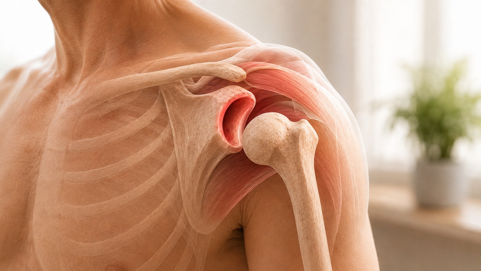
The shoulder is a ball-and-socket joint. The ball at the top of the upper arm bone, called the humeral head, sits within a shallow socket called the glenoid.
The shoulder socket is much shallower than the hip socket. This gives the arm an enormous range of movement, but it also means the joint relies heavily on soft tissues and muscles to remain stable. The main stabilising structures include the labrum (a rim of cartilage around the socket), the joint capsule, the shoulder ligaments, the rotator cuff muscles, the muscles that control the shoulder blade, and the nervous system, which coordinates movement and joint position.
Shoulder instability means that the ball moves excessively within the socket and produces symptoms such as pain, slipping, apprehension, subluxation or complete dislocation.
Dislocation, subluxation and instability: what is the difference?
Shoulder dislocation
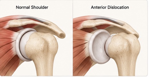
A dislocation occurs when the ball comes completely out of the socket. The shoulder may look visibly deformed, movement becomes extremely painful and the arm may feel locked in one position. Most complete dislocations need to be put back into place by a trained healthcare professional.
Shoulder subluxation
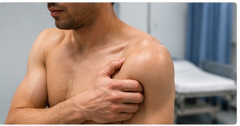
A subluxation is a partial dislocation. The ball moves partly out of the socket but returns by itself or with a change in position. People often describe this as “it slipped out and back in”, “it felt as though the shoulder moved”, “it nearly came out” or “I felt a clunk”.
Shoulder instability
Instability is the wider term. A person may have instability even if the shoulder has not fully dislocated again. They may feel that it is about to come out during certain movements, particularly when the arm is raised and rotated backwards. This sensation is called apprehension.
What should you do if the shoulder is currently dislocated?
A suspected shoulder dislocation requires urgent medical assessment. Do not ask an untrained person to pull the shoulder back into place. A dislocation may be accompanied by a fracture, nerve injury, blood vessel injury, rotator cuff damage or injury to the cartilage and ligaments.
The shoulder should be assessed, X-rayed where appropriate and reduced in a controlled environment with suitable pain relief and muscle relaxation. The nerves and circulation in the arm should be checked before and after reduction, and a further X-ray is normally used to confirm that the joint is back in place.
Seek urgent medical attention if the shoulder looks deformed, movement is impossible, or there is numbness, weakness, a cold hand or loss of pulse.
Which way does the shoulder dislocate?
Anterior shoulder dislocation
More than 95% of traumatic shoulder dislocations occur in an anterior direction, meaning the ball moves forwards out of the socket. This commonly happens when the arm is lifted away from the body, rotated outwards and forced backwards. Typical causes include falling onto an outstretched hand, a rugby tackle, a fall while skiing or cycling, contact sport and a forceful overhead movement.
Posterior shoulder dislocation
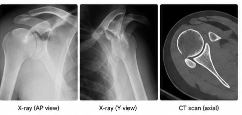
Posterior dislocation is much less common. The ball moves backwards and may be difficult to recognise because the shoulder does not always look dramatically deformed. It can occur after a seizure, an electric shock, a fall onto an internally rotated arm or a strong direct force to the front of the shoulder. Because posterior dislocations can be missed on a standard front-view X-ray, an additional side or axial view is important when the diagnosis is suspected.
Inferior shoulder dislocation
Inferior dislocation is rare. The arm is often held fixed above the head and requires emergency treatment.
What happens inside the shoulder during a dislocation?
A traumatic shoulder dislocation may damage several structures at the same time. Understanding these injuries helps explain why one person may recover with physiotherapy while another may benefit from surgery.
Bankart lesion
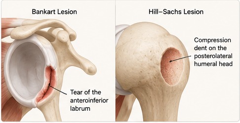
The labrum acts like a raised bumper around the edge of the socket. During an anterior dislocation, the ball may peel this bumper away from the front and lower part of the socket. This is called a Bankart lesion. Once the labrum has detached, the socket may no longer hold the ball as securely, and the capsule and ligaments may also become stretched. A Bankart lesion is one of the commonest structural causes of recurrent anterior shoulder instability.
Bony Bankart lesion
Sometimes the force of the dislocation pulls away not only the labrum but also a fragment of bone from the front of the socket. This is called a bony Bankart lesion. Repeated dislocations may gradually wear away more of the socket, rather like repeated impacts chipping the edge of a shallow bowl.
Hill-Sachs lesion
As the ball comes out of the socket, the harder edge of the socket can create a dent in the humeral head. This is known as a Hill-Sachs lesion. A small dent may have little effect. A larger defect, or one positioned so that it catches on the edge of the socket, can increase the risk of the shoulder dislocating again.
Rotator cuff tear
Rotator cuff tears are particularly important in people over 40. A younger person is more likely to tear the labrum and remain unstable. An older person is less likely to dislocate repeatedly but more likely to tear the rotator cuff during the original injury. Persistent weakness or inability to raise the arm after a dislocation may therefore indicate a rotator cuff tear rather than simple post-injury pain.
Nerve injury
The axillary nerve runs close to the shoulder joint and may be stretched during a dislocation. This can cause numbness over the outer shoulder, weakness of the deltoid muscle and difficulty lifting the arm. Most nerve injuries recover, but persistent numbness or weakness requires review.
Fractures
A shoulder dislocation may also cause a fracture, particularly a greater tuberosity fracture, a glenoid rim fracture or a fracture of the humeral head or neck. The likelihood of an associated fracture increases with age and with higher-energy injuries.
Why do some people dislocate once while others keep dislocating?
This is the central question after a first shoulder dislocation. The answer depends on a combination of age, sex, activity level, sport, joint laxity, the damage caused by the first dislocation, whether instability symptoms continue, bone loss, and the person's goals and willingness to accept future risk. No single factor decides treatment on its own.
Age is one of the strongest predictors
The younger a person is at the time of the first traumatic dislocation, the greater the chance of recurrent instability. Teenage males are at particularly high risk. Estimates from one large study suggested that males around the age of 20 had a recurrence risk of approximately 72% within the first two years, with the risk reducing progressively as age increased.
A broader evidence review found that people aged 40 or younger were substantially more likely to experience recurrence than older patients. Men and people with hyperlaxity were also at increased risk. This does not mean that every young person should automatically have surgery — it means that the risk of recurrence needs to be discussed seriously.
Sport and occupation matter
The shoulder is exposed to more risk during activities involving tackling, collisions, throwing, overhead movement, falling, forceful lifting and working with the arm above shoulder height. A young rugby player who wants to return to contact sport has a different risk profile from someone who does not take part in contact or overhead activities. Surgery may therefore be reasonable after a first dislocation for one patient but unnecessary for another person of the same age.
The number of instability episodes matters
A shoulder that has dislocated several times has already demonstrated that it is not reliably stable. Repeated episodes can lead to further labral damage, stretching of the capsule, increasing socket bone loss, enlargement of a Hill-Sachs lesion, cartilage damage, reduced confidence and difficulty returning to sport or work.
For this reason, recurrent dislocation makes surgery much more likely. The decision is not based only on the number of episodes: a single further subluxation during light daily activity may be more concerning than a brief sensation of uncertainty during a very high-risk sporting movement.
Symptoms between dislocations matter
Some people have one dislocation and later feel completely normal. Others develop persistent symptoms such as the shoulder slipping, fear when the arm is overhead, inability to throw, avoiding certain positions, pain and weakness, recurrent subluxations and loss of confidence in sport or work. A shoulder does not need to dislocate repeatedly before it becomes a significant problem — persistent apprehension can be disabling even when the ball has not fully come out again.
Joint laxity and hypermobility
Some people naturally have more flexible joints. Joint laxity is not automatically a medical problem; many people are flexible but have no pain or instability. Laxity is a physical finding, while instability is a symptom.
A person with naturally loose joints may rely more heavily on muscle control to keep the shoulder centred. If those muscles are weak, poorly coordinated or fatigued, the shoulder may begin to sublux without a major injury. This is known as atraumatic shoulder instability.
First-time shoulder dislocation: do you always need surgery?
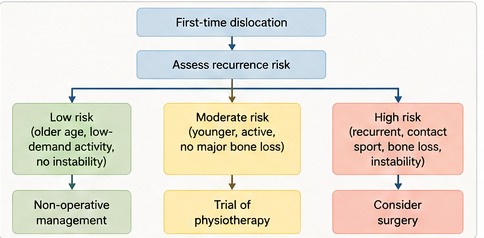
No. Many first-time dislocations can be treated successfully without an operation. However, the decision should not simply be “first dislocation equals physiotherapy” or “young person equals surgery”. A good decision considers the person as a whole.
When non-surgical treatment may be reasonable
Non-surgical treatment may be appropriate when this is the first dislocation, the person has a relatively low risk of recurrence, the shoulder feels stable after recovery, there is no major socket bone loss, there is no large or clinically important associated injury, the person does not take part in high-risk contact or overhead activity, they would prefer to avoid surgery, and they understand and accept the possibility of another dislocation.
A typical example might be an older adult who dislocated the shoulder after a fall, regained movement and strength and has no ongoing instability. That person may gain little from immediate stabilisation surgery.
When surgery may be discussed after a first dislocation
Surgery may be discussed earlier when several high-risk factors are present, including teenage or young adult age, male sex, contact or collision sport, high-level overhead sport, an occupation with substantial overhead or physical demands, significant labral injury, socket bone loss, a clinically important Hill-Sachs lesion, strong concern about recurrence and a need to return reliably to high-risk activity.
Evidence suggests that surgery reduces recurrent instability in young, active patients compared with non-operative treatment. However, surgery also introduces risks and requires months of rehabilitation, so shared decision-making is essential.
Why surgeons do not simply operate on every first dislocation
Not every shoulder will dislocate again: even in younger groups recurrence is not inevitable, some labral injuries scar into a stable position and some people regain excellent control with rehabilitation.
Surgery also has risks, including infection, stiffness, nerve injury, persistent pain, recurrent instability, loss of external rotation, problems with anchors or grafts and the need for further surgery. Recovery is prolonged — a stabilisation operation is not a quick way back to sport.
Individual goals differ. A person returning to elite rugby may accept the risks of surgery to reduce the chance of recurrence, while someone with lower physical demands may prefer rehabilitation and accept that surgery could be considered later if instability returns.
Why surgeons do not simply wait for everyone to dislocate repeatedly
Repeated dislocations are not always harmless. Each episode may add further damage to the labrum, the ligaments, the socket bone, the humeral head and the joint cartilage. Once substantial bone loss has developed, a straightforward soft-tissue repair may be less likely to work and the operation required may then be more complex. This is why early surgery may be considered in a young, high-risk athlete even after the first dislocation.
A practical decision pathway after a traumatic dislocation
Step 1 — Confirm that the shoulder has been reduced safely. The shoulder should be examined and imaged to confirm its position and identify associated fractures.
Step 2 — Check for associated injury: nerve function, circulation, rotator cuff strength, fractures and persistent inability to raise the arm.
Step 3 — Estimate the risk of recurrence using age, sex, sport, occupation, hyperlaxity, the mechanism of injury, imaging findings and personal goals.
Step 4 — Begin early recovery. A sling may be used for comfort, and gentle movement usually begins once pain allows and according to the advice of the treating team. Prolonged immobilisation beyond one week has not been shown to reduce recurrence in younger patients, although the duration should be adapted when a fracture or other injury is present.
Step 5 — Reassess stability and function: does the shoulder feel secure, can the arm be raised, is strength returning, is there apprehension, has it slipped again, can you return to work or sport?
Step 6 — Decide between continued rehabilitation and surgery, based on risk, symptoms, scans, activity requirements and patient preference rather than on one MRI finding.
What does physiotherapy involve after a shoulder dislocation?
Physiotherapy aims to restore comfortable movement, rotator cuff strength, shoulder blade control, joint position awareness, confidence, tolerance of everyday activity and sport-specific function.
Early rehabilitation may include hand, wrist and elbow movement, gentle assisted shoulder movement, posture and shoulder blade exercises and isometric muscle activation. Later stages may include progressive rotator cuff strengthening, resistance-band exercises, controlled overhead movement, pushing and pulling exercises, balance and joint-position training, throwing or contact preparation and sport-specific drills.
Rehabilitation is not only about building muscle. It also retrains how the brain and shoulder muscles respond when the arm moves into positions that previously felt unsafe.

How long does recovery take without surgery?
Recovery varies according to pain, age, associated injury, occupation, sport, strength, confidence and response to rehabilitation. Movement commonly improves over the first several weeks, while strength and confidence may take several months.
Return to contact sport should not be based only on the passage of time. The shoulder should have near-normal movement, good strength, no significant apprehension, satisfactory functional control and the ability to complete sport-specific tasks.
What is atraumatic shoulder instability?
Not every unstable shoulder begins with a major injury. Atraumatic shoulder instability develops without a clear traumatic event. The shoulder may sublux during ordinary movements, feel insecure when reaching, move excessively during sport, slip while sleeping, feel unstable in more than one direction or dislocate through abnormal muscle activation.
This type of instability often affects younger people and may be associated with naturally loose joints, repetitive overhead activity, reduced rotator cuff control, poor shoulder blade control, altered movement patterns, deconditioning, fear and avoidance of movement, and complex pain or neurological symptoms.
Some people can initially make the shoulder move out deliberately. Repeatedly doing this as a “party trick” is discouraged because the movement may become difficult to control.
How is atraumatic instability treated?
Specialist physiotherapy is the main treatment. The programme may focus on improving rotator cuff coordination, retraining shoulder blade movement, restoring joint position awareness, correcting abnormal muscle patterns, strengthening the wider kinetic chain, gradually rebuilding confidence and returning to meaningful activities.
Improvement can take several months. Reported outcomes vary, but many patients improve without surgery. Specialist guidance recommends allowing a structured programme enough time to work, with reassessment after approximately 12 weeks and continued treatment where progress is being made.
Why is surgery approached cautiously in atraumatic instability?
In traumatic instability there is often a clear structural target such as a detached labrum. In atraumatic instability the shoulder may be structurally normal and the main problem may be altered muscle control. Tightening or repairing the shoulder without addressing that control problem may fail to resolve symptoms, create stiffness, worsen pain, lead to recurrent instability or result in further surgery.
Surgery is therefore usually considered only when high-quality specialist rehabilitation has been completed, symptoms remain severe, there is a clearly identified structural problem and surgical target, the patient understands the substantial risk of failure, and the case has been assessed by an experienced shoulder team.
How is shoulder instability diagnosed?
Diagnosis starts with the history. Important questions include whether there was a clear injury, whether the shoulder was completely out or slipped partly, whether it needed reduction in hospital, how many episodes have occurred, whether it happens during sport or ordinary activity, whether the shoulder feels unstable at rest or during sleep, whether other joints are unusually flexible, whether there is numbness or weakness, what activities have been stopped and what the person's goals are.
The examination may assess range of movement, rotator cuff strength, shoulder blade movement, joint laxity, apprehension, nerve function, generalised hypermobility and the direction of instability. The feeling that the shoulder may dislocate is often more informative than pain alone during instability testing.
Which scans are used?
X-rays are the first investigation for an acute dislocation. They help confirm the diagnosis, show the direction of dislocation, identify fractures and confirm that the shoulder is back in place. More than one view is important because a posterior dislocation may be missed on a single front-view X-ray.
Ultrasound may be used when a rotator cuff tear is suspected, particularly in older patients with persistent weakness after the shoulder has been reduced.
MRI shows soft tissues including the labrum, capsule, rotator cuff, cartilage, ligaments and biceps tendon. An MRI arthrogram involves injecting contrast into the joint before the scan, which may make subtle labral or capsular injuries easier to see.
CT is particularly useful for assessing bone. It can measure socket bone loss, a bony Bankart lesion, the size and position of a Hill-Sachs defect and the shape of the glenoid. In short: MRI mainly shows what has happened to the soft tissues, while CT is often better for showing how much bone has been lost.
Does every Bankart lesion need surgery?
No. A Bankart lesion indicates that the labrum has detached from the front of the socket, but treatment still depends on age, activity level, recurrence risk, instability symptoms, bone loss and patient preference.
A low-risk patient who becomes stable after one dislocation may not need the labrum repaired. A young contact athlete with repeated instability and little bone loss is much more likely to benefit from a Bankart repair.
What is an arthroscopic Bankart repair?
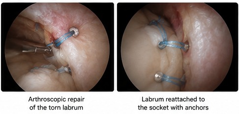
An arthroscopic Bankart repair is a keyhole stabilisation operation. A camera is inserted into the shoulder, the detached labrum is identified and the edge of the socket is prepared. Small anchors are inserted into the bone, sutures from the anchors are passed through the labrum and capsule, and the labrum is reattached to the socket. The stretched capsule may also be tightened.
The aim is to restore the cartilage bumper and tighten the soft tissues that hold the ball in the socket. Modern arthroscopic repair using suture anchors has outcomes comparable with open anatomical repair when there is no major bone defect.
Who is most suited to a Bankart repair?
A Bankart repair is most suitable when instability is mainly anterior, the labrum is detached, the capsule is stretched, socket bone loss is limited, the Hill-Sachs lesion is unlikely to engage significantly, there has not been a failed previous repair and the patient can complete rehabilitation. It may be less reliable when there is substantial bone loss or a large, unfavourably positioned Hill-Sachs lesion.
What is remplissage?
Remplissage is a French word meaning “to fill”. It is an additional keyhole procedure that may be combined with a Bankart repair when a Hill-Sachs lesion is likely to catch on the edge of the socket. The surgeon attaches part of the posterior capsule and infraspinatus tendon into the defect, making the dent less able to engage with the front edge of the socket during movement.
Remplissage may reduce recurrence in selected patients, although it can reduce external rotation slightly and the precise indications continue to evolve.
What is the Latarjet procedure?
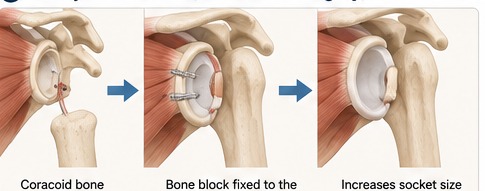
The Latarjet procedure is used when simply repairing the labrum may not provide enough stability, often because the front of the socket has lost a significant amount of bone. A piece of bone called the coracoid is detached from the shoulder blade and transferred to the front of the glenoid, where screws hold the bone block in place. The attached tendons provide an additional sling effect when the arm moves into a vulnerable position.
A simple way to understand this is that a Bankart repair restores the damaged soft-tissue bumper, while a Latarjet rebuilds and reinforces the front of a socket that has become too small or damaged.
Why not perform a Latarjet on everyone?
The Latarjet can provide excellent stability, but it is a larger operation than a straightforward Bankart repair. Potential complications include graft fracture, failure of the bone block to unite, screw problems, nerve injury, infection, stiffness, arthritis and recurrent instability.
It is therefore generally reserved for patients likely to benefit from the added bony stability, including those with significant glenoid bone loss, failed previous soft-tissue stabilisation, selected high-risk recurrent instability and certain combinations of socket and humeral-head bone loss. Older guidance used around 20% glenoid bone loss as an important threshold, although more recent work suggests that smaller amounts of “subcritical” bone loss may also reduce the success of an isolated Bankart repair.
How does a surgeon choose the operation?
The operation is chosen by answering several questions. Is the instability traumatic or atraumatic? A traumatic labral tear may have a clear surgical target, while atraumatic instability is usually treated with muscle retraining first.
Is the instability anterior, posterior or in more than one direction? The direction determines which part of the capsule and labrum requires treatment.
How much socket bone has been lost? Limited bone loss may be compatible with a Bankart repair; more substantial bone loss may require a bone-block procedure.
Is there a significant Hill-Sachs lesion? A clinically important defect may require remplissage or influence the choice of a Latarjet procedure.
Has surgery already failed? A failed previous Bankart repair raises concern about missed bone loss, poor tissue quality, hyperlaxity, an off-track Hill-Sachs lesion, ongoing high-risk activity or an incorrect original diagnosis.
Finally, what does the patient need the shoulder to do — contact sport, throwing, swimming, heavy manual work, military duties or ordinary daily activity?
| Treatment | Most suitable for | Main advantage | Important limitation |
|---|---|---|---|
| Physiotherapy after traumatic dislocation | First-time dislocation with acceptable recurrence risk | Avoids surgery and restores movement and strength | The shoulder may dislocate again |
| Specialist physiotherapy for atraumatic instability | Instability without a major injury, hyperlaxity or altered muscle control | Treats the underlying movement problem | Requires time, engagement and specialist expertise |
| Arthroscopic Bankart repair | Traumatic anterior instability with limited bone loss | Repairs the labrum through keyhole surgery | Less reliable when significant bone loss is present |
| Bankart repair with remplissage | Bankart lesion with a clinically important Hill-Sachs defect | Addresses both labral damage and humeral-head engagement | May reduce external rotation |
| Latarjet procedure | Significant socket bone loss, failed repair or selected high-risk instability | Rebuilds the socket and adds dynamic stability | Larger operation with graft, screw and nerve-related risks |
| Capsular shift | Selected atraumatic or multidirectional instability with a clear structural indication | Tightens an excessively loose capsule | Evidence is limited and patient selection is critical |
Risks of shoulder stabilisation surgery
Possible complications include infection, stiffness, persistent pain, nerve injury, blood vessel injury, recurrent instability, loss of movement, failure of the labrum to heal, graft or screw problems after bone-block surgery, later arthritis and the need for revision surgery.
The likelihood of recurrence differs between operations and patient groups. A review reported pooled recurrence rates of approximately 16% after labral repair, 9% after labral repair with remplissage and 6% after bone augmentation. These figures came mainly from patients treated for recurrent instability and should not be treated as an individual prediction. Bone augmentation also carried a higher overall complication rate than labral repair.
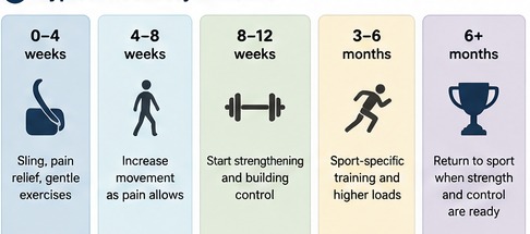
Recovery after shoulder stabilisation surgery
Recovery depends on the operation performed, tissue and bone healing, the surgeon's protocol, whether the procedure is primary or revision surgery, and sport or work demands.
| Time after surgery | Typical stage |
|---|---|
| 0–4 weeks | Sling protection, wound care, hand and elbow movement, prescribed shoulder exercises |
| 4–8 weeks | Gradual increase in shoulder movement |
| 8–12 weeks | Progression of active movement and early strengthening |
| 3–4 months | More advanced strengthening and functional rehabilitation |
| 4–6 months | Gradual return to heavier work and non-contact sporting activity |
| Around 6 months or later | Possible return to contact sport after strength and functional testing |
| 6–12 months | Continued improvement in confidence, endurance and performance |
When can you drive?
You should not drive while wearing a sling. Before returning to driving you should be able to control the steering wheel, use the gears and handbrake, perform an emergency manoeuvre, react quickly without hesitation and comply with your insurer's requirements. Your surgical team should advise when this is safe.
When can you return to sport?
Return to sport should be based on recovery rather than time alone. Before returning, the shoulder should usually have satisfactory movement, near-normal strength, good control, no significant apprehension, the ability to tolerate sport-specific drills and confidence during vulnerable positions.
Contact athletes need to tolerate tackling and impact preparation before returning to competition. Throwing athletes may need a staged throwing programme, because small losses of rotation or control can have a large effect on performance.
Can shoulder instability cause arthritis?
A shoulder that has dislocated is at increased risk of developing arthritis later in life. The risk appears to be influenced by the original injury, repeated dislocations, bone and cartilage damage, age at the first dislocation, high-energy sport, and surgery and the type of procedure performed.
Long-term studies show that recurrent instability is associated with greater joint damage, but surgery does not guarantee that arthritis will be prevented. The main aim of treatment is therefore to restore stability, function and confidence while reducing further injury where possible.
When should you seek advice?
Arrange a specialist assessment if the shoulder has dislocated more than once, it repeatedly subluxes or slips, you avoid overhead positions because it feels unstable, you cannot return to work or sport, symptoms continue despite physiotherapy, you are a young athlete considering return to contact sport, weakness persists after the dislocation, there is continuing numbness, previous stabilisation surgery has failed, the shoulder dislocates during low-energy activities or at rest, or atraumatic instability is causing major disruption to school, work or daily life.
Seek urgent medical attention if the shoulder is currently out of joint, the arm looks deformed, the hand is cold, pale or blue, the pulse feels reduced, there is new or worsening numbness, the arm cannot be moved after a significant injury, there is severe swelling, the joint is hot or red, you have a fever or feel generally unwell, shoulder pain follows a seizure or electric shock, or there is chest pain, shortness of breath or clamminess.
Top tips
- Do not try to force a dislocated shoulder back yourself — associated fractures and nerve injuries must be considered first.
- Understand your personal recurrence risk: age, sport, hyperlaxity, repeated episodes and bone loss matter more than the fact that a dislocation happened.
- Do not judge recovery by pain alone — strength, confidence, movement and the feeling of stability are all important.
- Give specialist physiotherapy a proper chance when instability is atraumatic; surgery cannot reliably correct a problem caused mainly by abnormal muscle control.
- Ask why a particular operation has been recommended — Bankart repair, remplissage and Latarjet solve different problems.
Related articles
Rotator Cuff Tear: Symptoms, Treatment, Surgery and Recovery
An evidence-based guide to rotator cuff tears: symptoms, diagnosis, physiotherapy, injections, surgery and recovery.
Shoulder Impingement: Symptoms, Exercises, Treatment and Recovery
An evidence-based guide to shoulder impingement (subacromial pain syndrome): symptoms, diagnosis, exercises, injections, surgery and recovery.